Dec. 6, 1850 - Hermann von Helmholtz demonstrated his revolutionary ophthalmoscope to the Berlin Physical Society.
Ophthalmoscopy, also called funduscopy, is a test that allows a health professional to see inside the fundus of the eye and other structures using an ophthalmoscope (or funduscope). It is done as part of an eye examination and may be done as part of a routine physical examination. It is crucial in determining the health of the retina, optic disc, and vitreous humor.


History
Although some credit the invention of the ophthalmoscope to Charles Babbage in 1847, it was not until it was independently reinvented by Hermann von Helmholtz that its usefulness was recognized - it was to revolutionize ophthalmology.
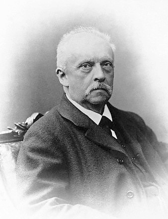 |
| Hermann von Helmholtz |
While training in France, Andreas Anagnostakis, an ophthalmologist from Greece, came up with the idea of making the instrument hand-held by adding a concave mirror. Austin Barnett created a model for Anagnostakis, which he used in his practice and subsequently when presented at the first Ophthalmological Conference in Brussels in 1857, the instrument became very popular among ophthalmologists.
In 1915, Francis A. Welch and William Noah Allyn invented the world's first hand-held direct illuminating ophthalmoscope, precursor to the device now used by clinicians around the world. This refinement and updating of von Helmholtz's invention enabled ophthalmoscopy to become one of the most ubiquitous medical screening techniques in the world today. The company Welch Allyn started as a result of this invention.
Medical Use
Ophthalmoscopy is done as part of a routine physical or complete eye examination. It is used to detect and evaluate symptoms of various retinal vascular diseases or eye diseases such as glaucoma.
In patients with headaches, the finding of swollen optic discs, or papilledema, on ophthalmoscopy is a key sign, as this indicates raised intracranial pressure (ICP) which could be due to hydrocephalus, benign intracranial hypertension or brain tumor, amongst other conditions. Cupped optic discs are seen in glaucoma.
In patients with diabetes mellitus, regular ophthalmoscopic eye examinations are important to screen for diabetic retinopathy as visual loss due to diabetes can be prevented by retinal laser treatment if retinopathy is spotted early.
In arterial hypertension, hypertensive changes of the retina closely mimic those in the brain, and may predict cerebrovascular accidents (strokes).
Etymology
The word ophthalmoscopy uses combining forms of ophthalmo- + -scopy, yielding "viewing the eye". The word funduscopy derives from fundus + -scopy, yielding "viewing the far inside".
No comments:
Post a Comment